Abstract
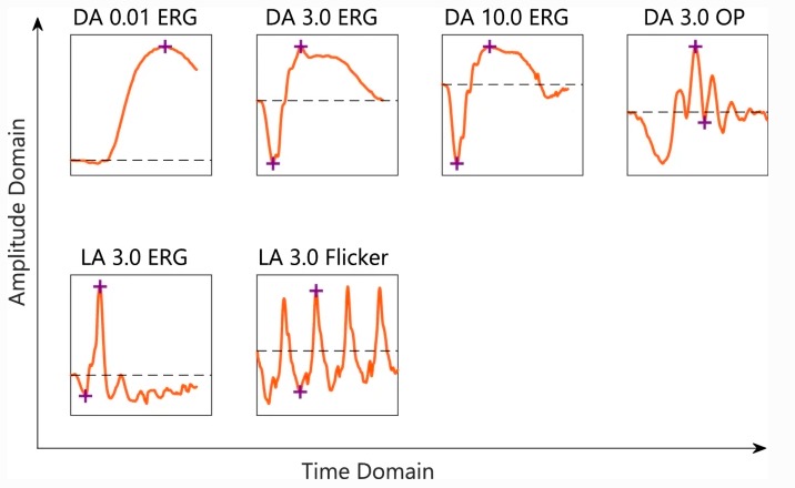
Purpose. Atropine eye drops are a common and effective treatment for slowing myopia progression, but the site and mode of action of atropine in controlling myopia are unclear. We investigated the early retinal sites of action of atropine by examining its effects on the human full-field electroretinogram (ffERG). Method. Baseline ffERGs were recorded in both eyes of 24 healthy subjects (mean +/- SD, 21.0 +/- 2.3 years; spherical equivalent refraction, range, + 1.63 to - 0.75 D) using 6 standard ISCEV protocols, 30 min after bilateral pupil dilation with 1% Tropicamide. Atropine (1 drop, 0.1%) was then instilled into the non-dominant eye. 24 h later, ffERGs were again recorded in both eyes. Ratios (post-atropine/ pre-atropine) of dark-adapted (DA) and light-adapted (LA) ffERGs were compared between atropine-treated and control eyes using multivariate repeated measures general linear models. Results. Atropine-treated eyes responded with 14% lower DA3.0 OP (oscillatory potential) amplitude (p = 0.003) and 4% delay in the DA10.0 a-wave peak time (p = 0.00099) compared with control eyes. Amplitudes and peak times were not different between atropine-treated and control eyes for DA0.01, LA3.0, and LA3.0 flicker ERGs. While atropine caused a small (1.26 mm2, p = 0.03) extra increase in pupil area in the treated eye, atropine-induced changes in ffERG responses bore no relationship with changes in pupil area (R2 = 2-5 percent, p > 0.05). Conclusions. The observed changes in oscillatory potentials corroborate previous findings that atropine affects neural activity in the inner retina. However, observed changes to the a-wave suggest that atropine also affects activity in photoreceptors.